Research methods 1Source of the documents on wood anatomy: this chapter and the one that follows were written using information and documents from the Laboratoire d’Anatomie at the Centre Technique du Bois, made available to us by C. Jacquiot, Research Director and Head of the Biology Department.
The scientific techniques used in this study were macroscopic examinations, microscopy and X-ray diffraction. 2The science of dendrochronology is part of current methodology in panel painting research. See Klein 1998a; 1998b, 2012; Daly 2007; Fraiture 2007, 2009; Bill et al. 2012. We made particular use of surface examination and microscopy to identify the wood, microscopy to examine the cloth and radiography to examine the preparation layers (ground). We also used chemical analysis as a supplementary research method in the identification of certain preparation layers. 3For coccolith analysis, see Perch-Nielsen et al. 2012; Stols-Witlox 2014.
Macroscopy and microscopy4Klein 2012: 51–65.
Wood identification
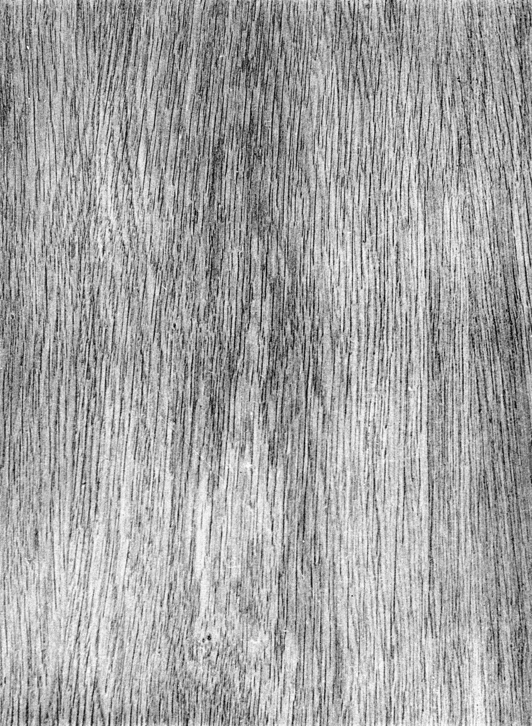
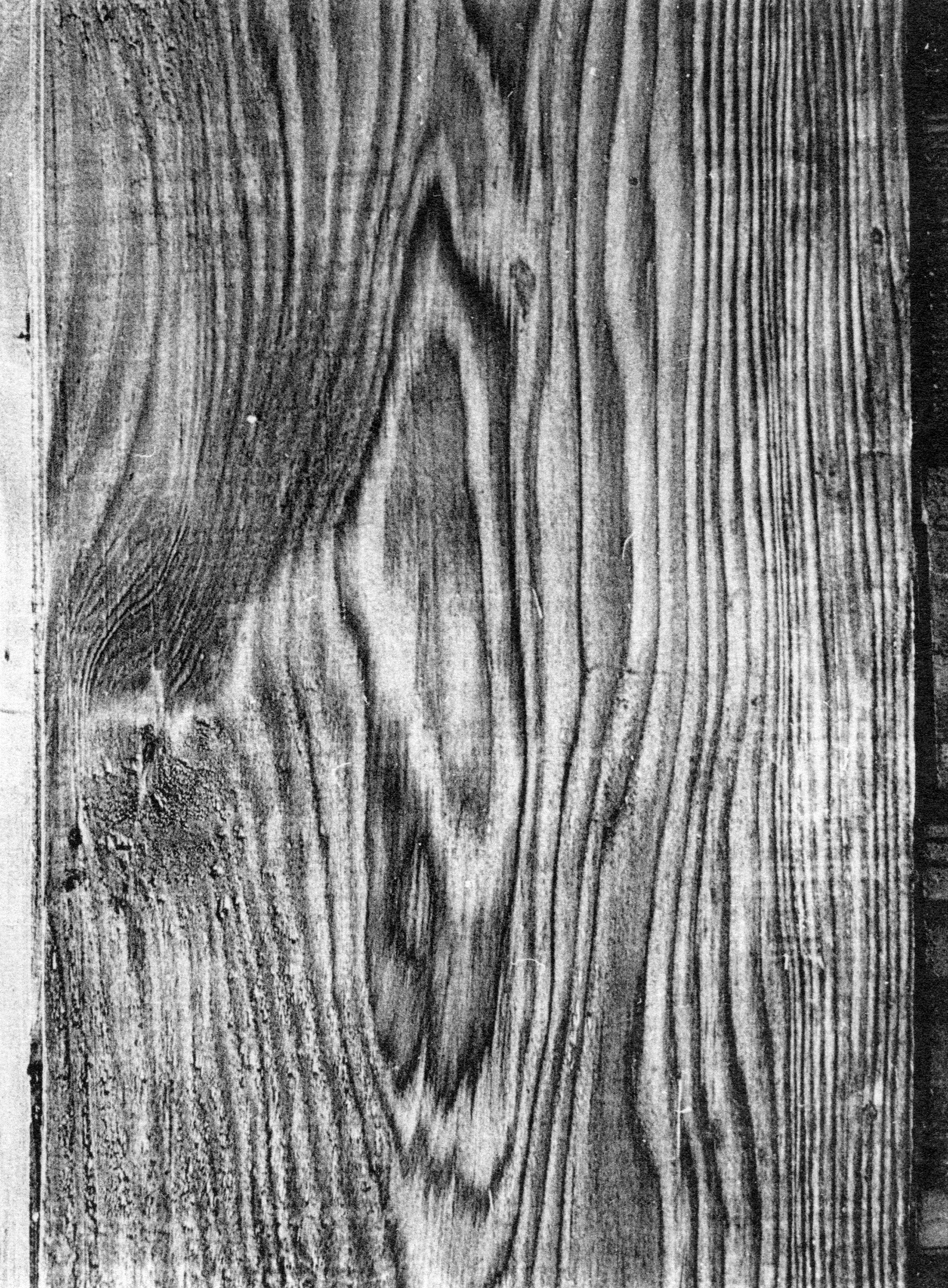
Where it is possible to view the backs of panels, the appearance of the wood – the presence of large vessels, medullary rays, wood grain, for instance – can provide an indication of its species. An area of surface exposed using a razor blade can then be observed with a magnifying glass (8× or 10×). The magnifying glass shows the structure of the wood in cross-section, revealing details such as the size of the rays, vessels and tyloses. It also enables us to identify, on a small sample, the grain of the wood and to take thin slices in the three principal planes. The slices are observed under the microscope, 5More detailed analysis requires use of a microscope. Microscopic examination begins at a magnification of 40, rising as high as 800 for the details. allowing examination of the details: the arrangement of the various tissues, and the characteristics of the fibres, vessels, rays, and pits. This type of observation can lead to the identification of the genus of the wood, and sometimes the species. (For different sections of a hardwood and softwood see Figs 30–33 below.)
Microscopy of wood species used by painters
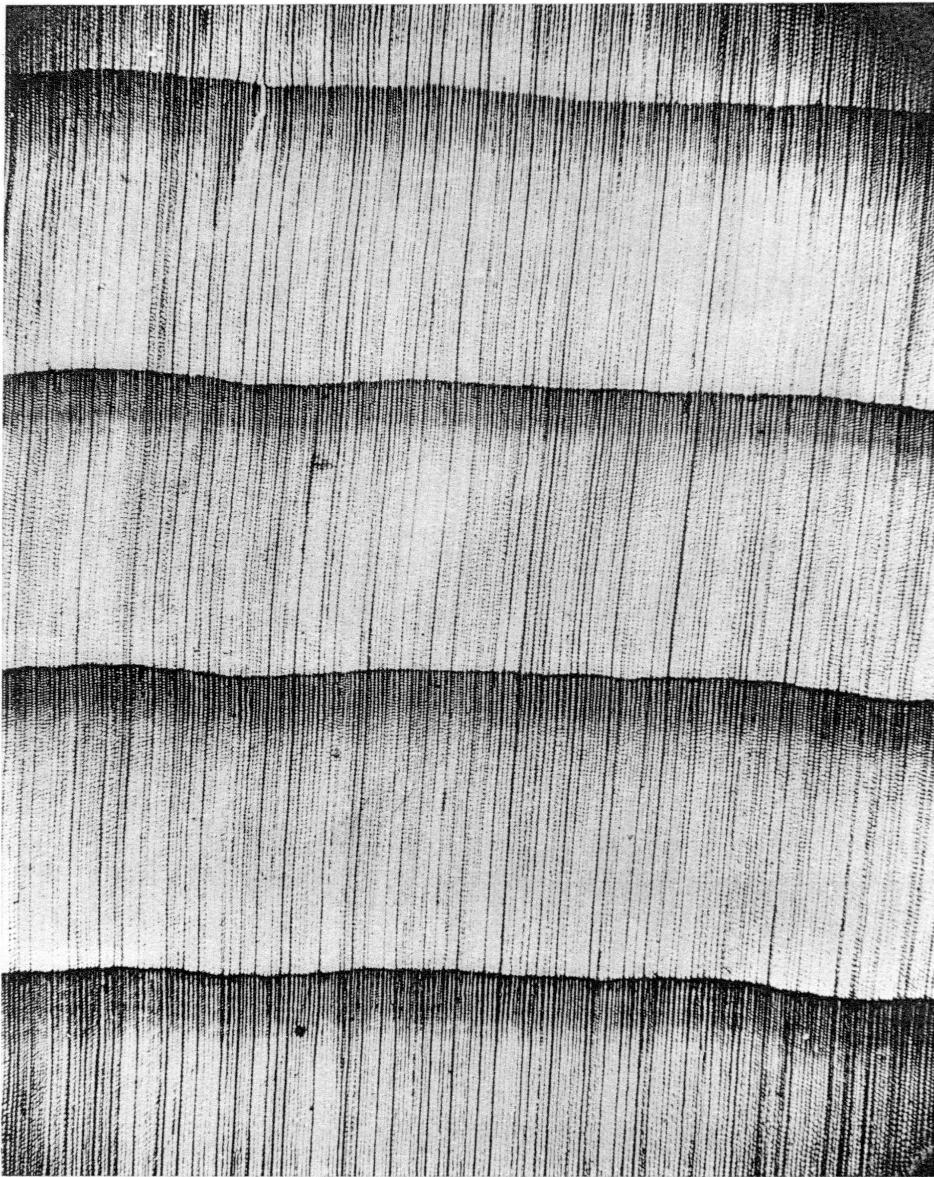
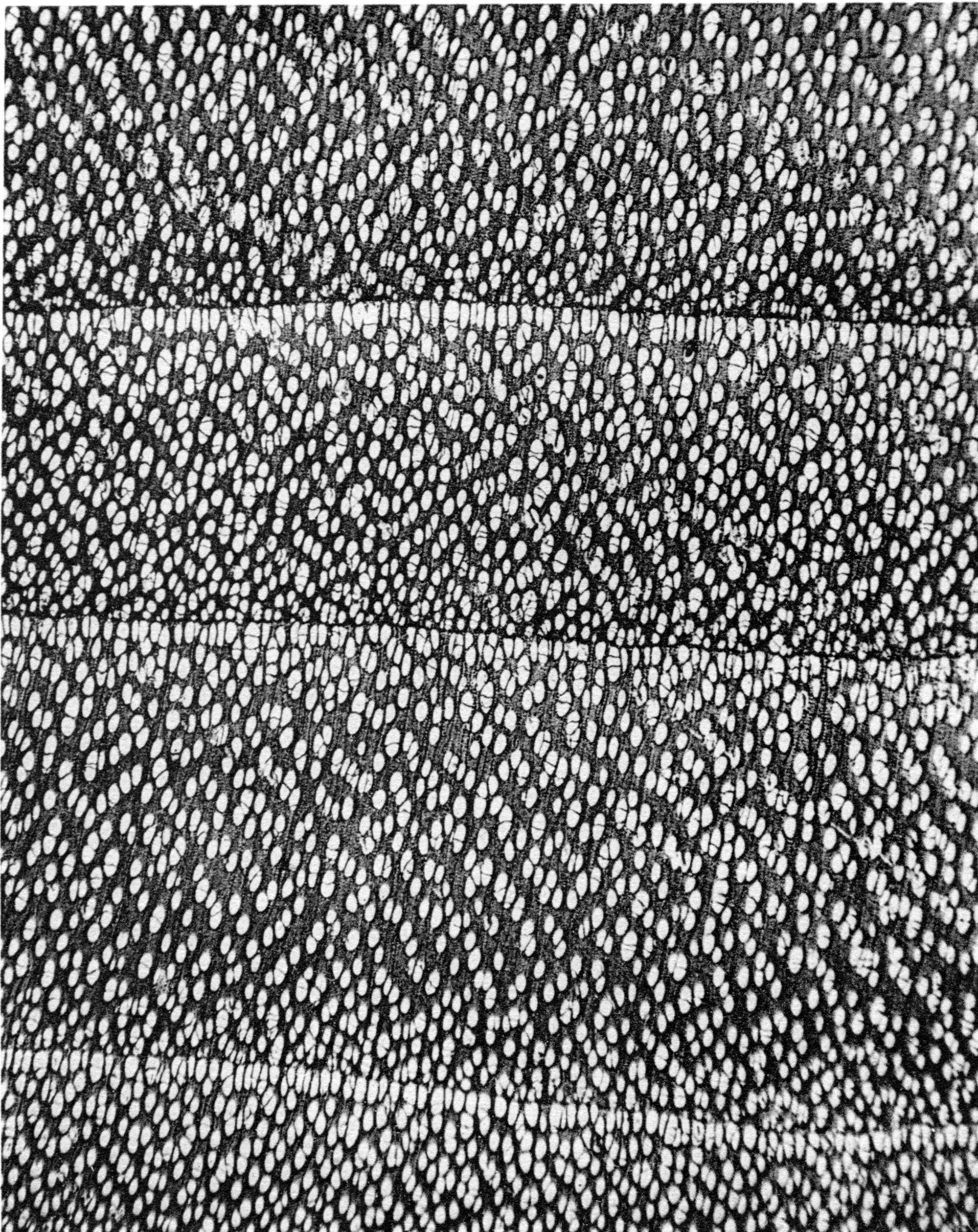
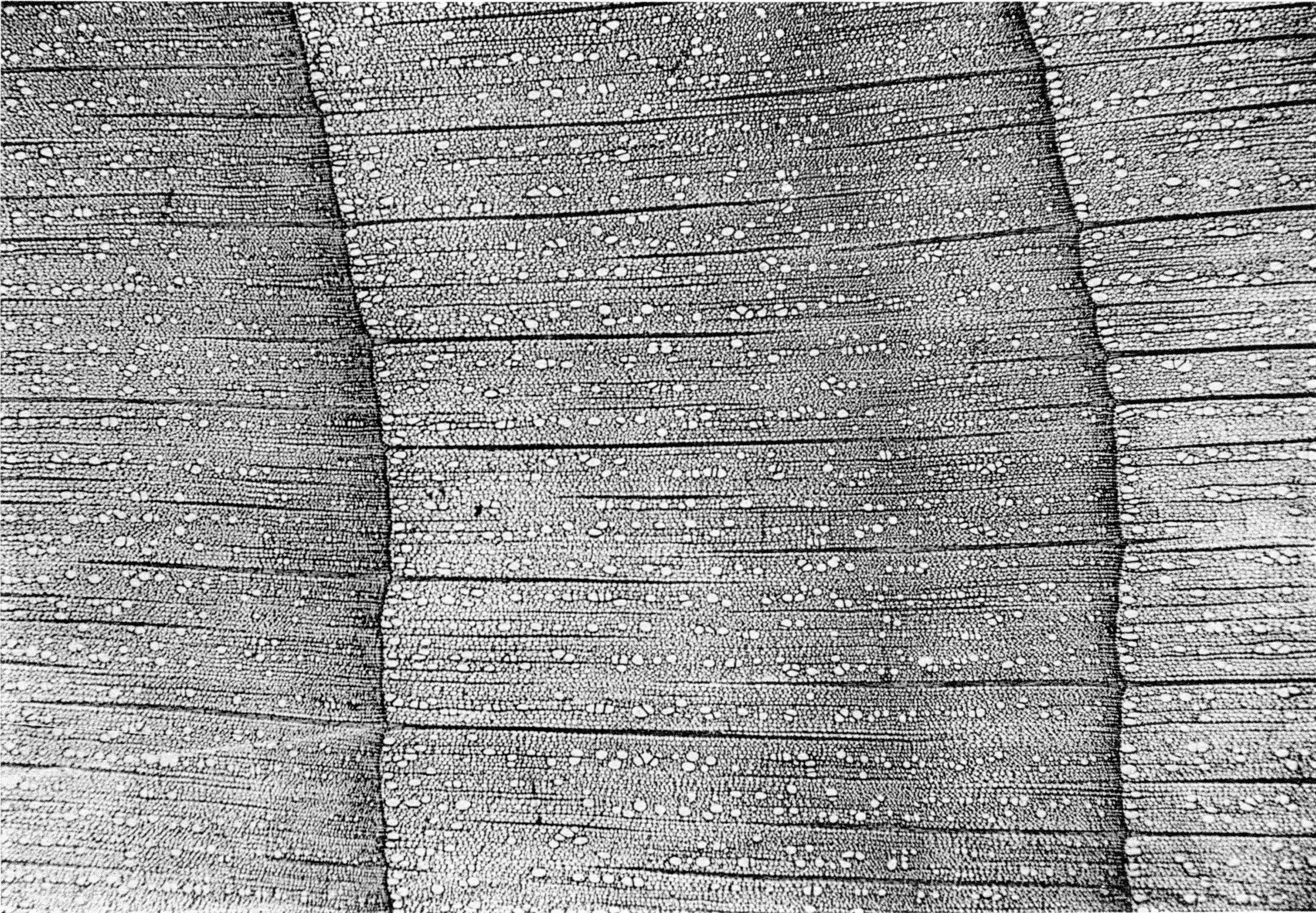
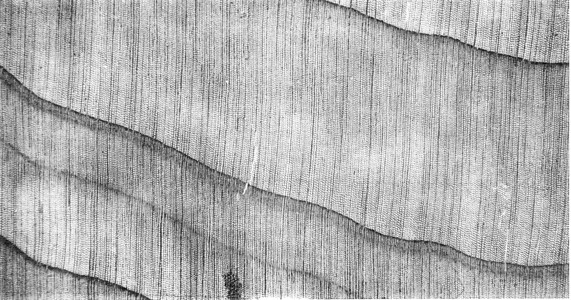
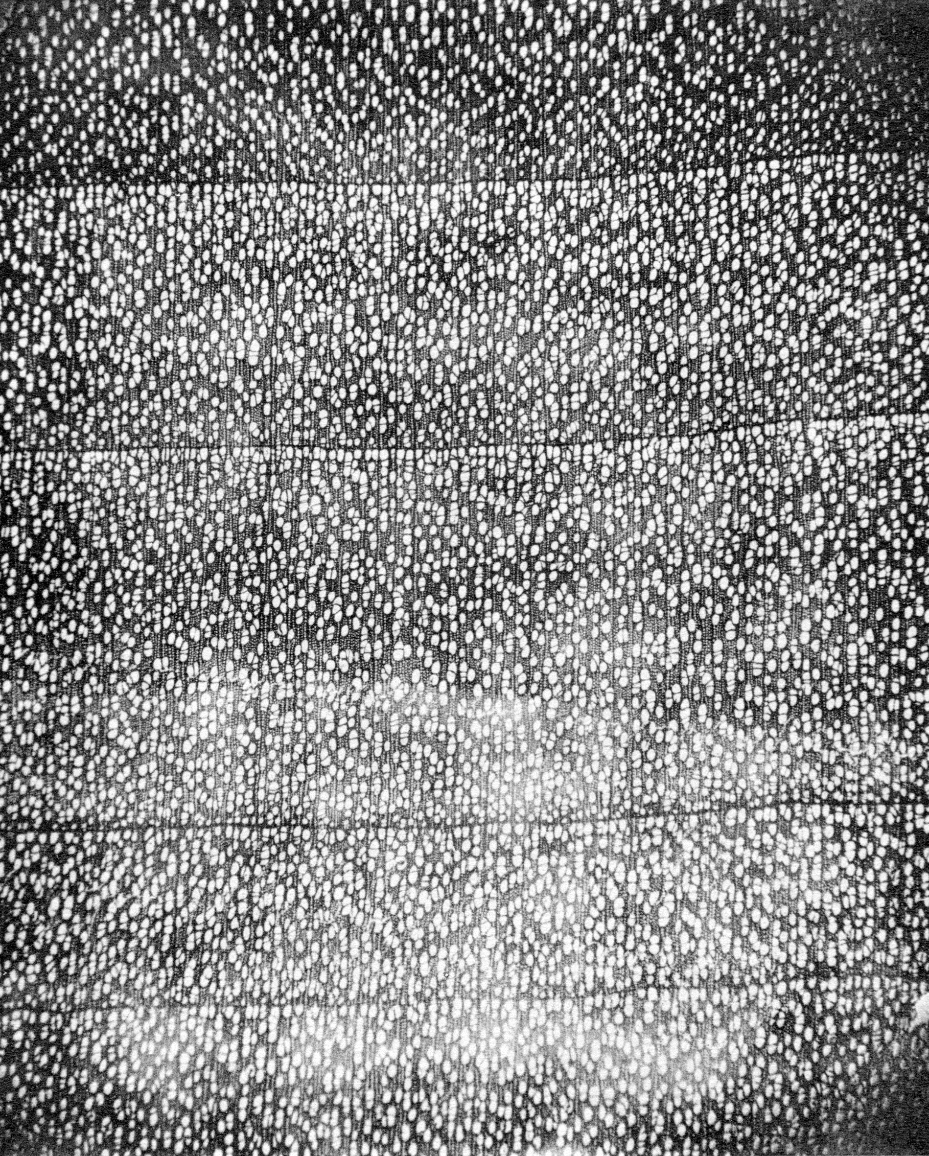
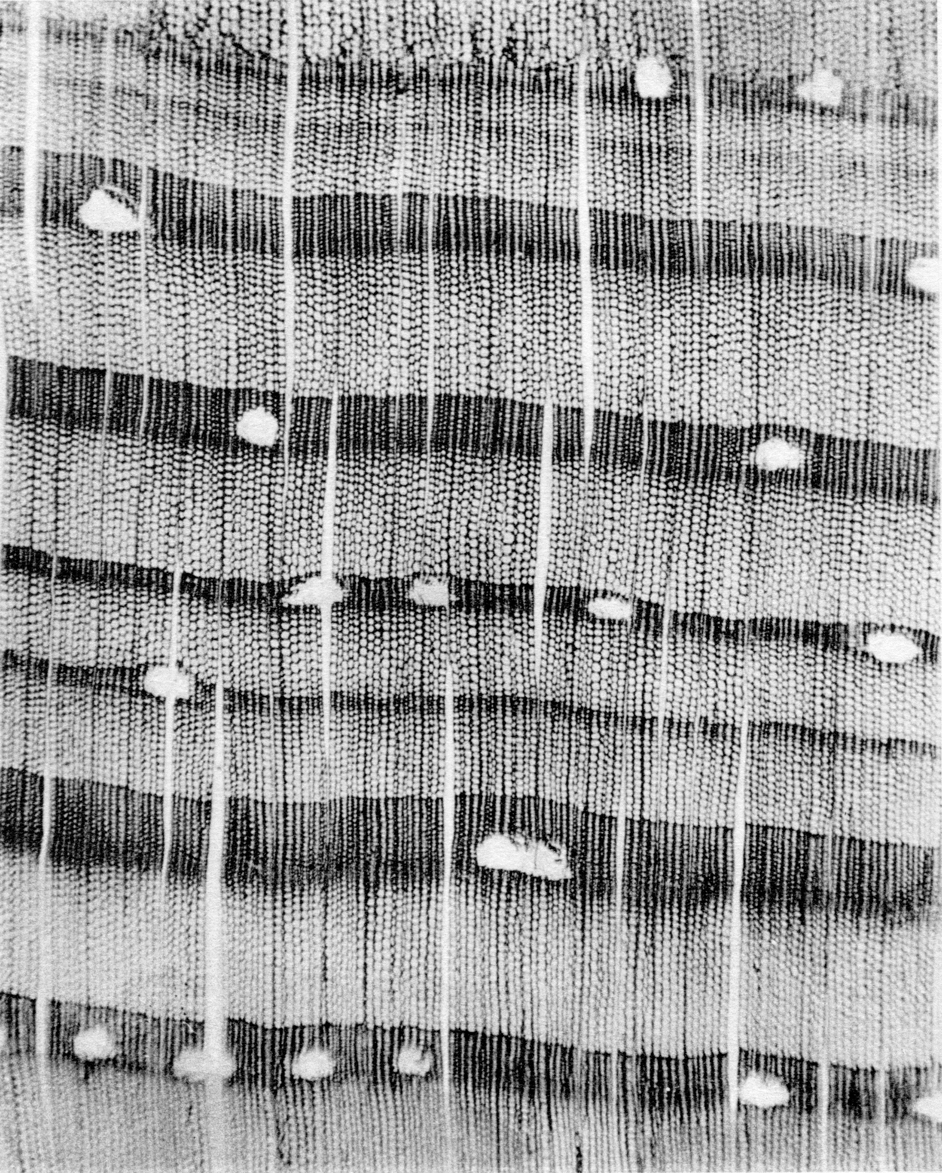
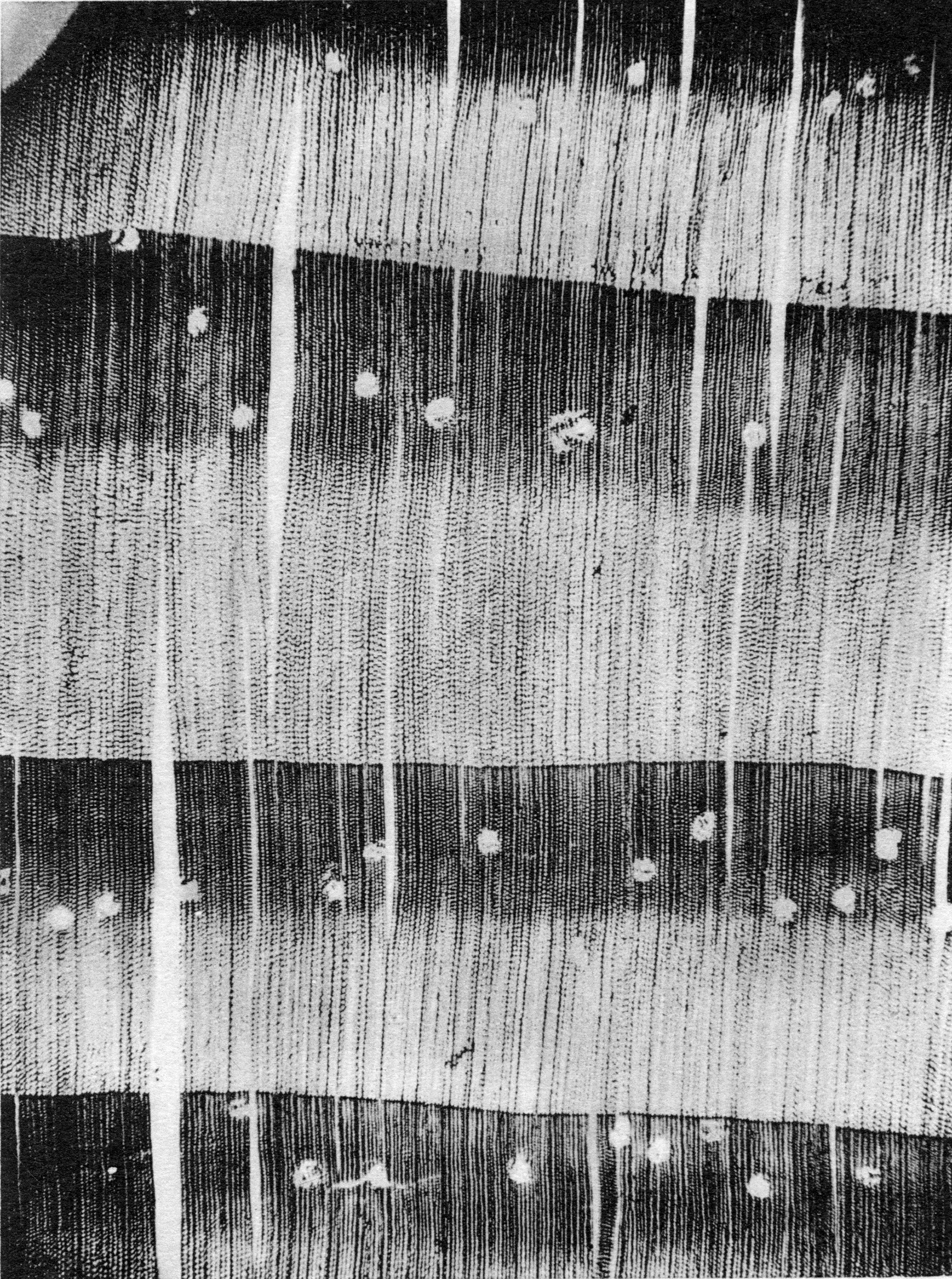
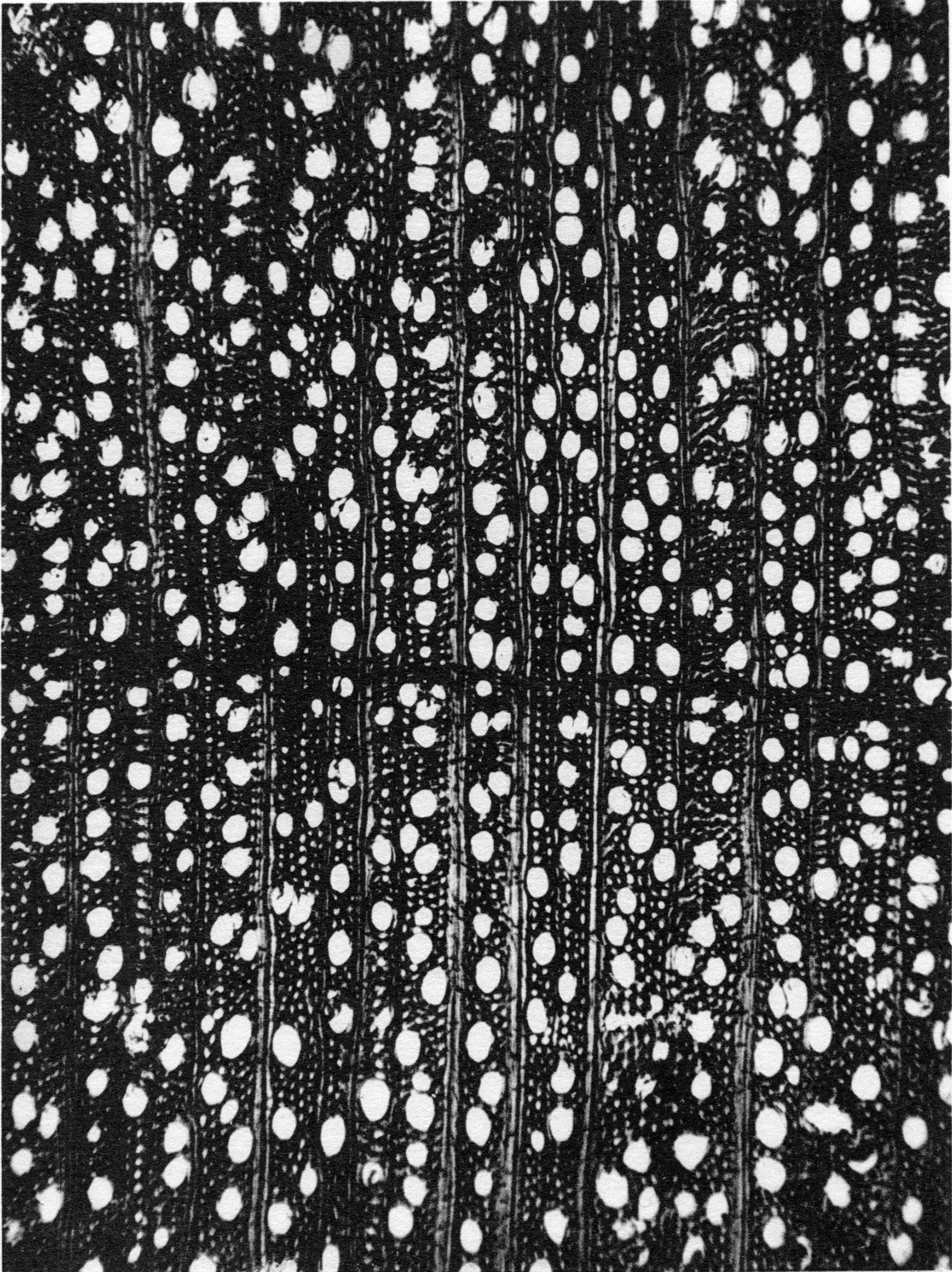
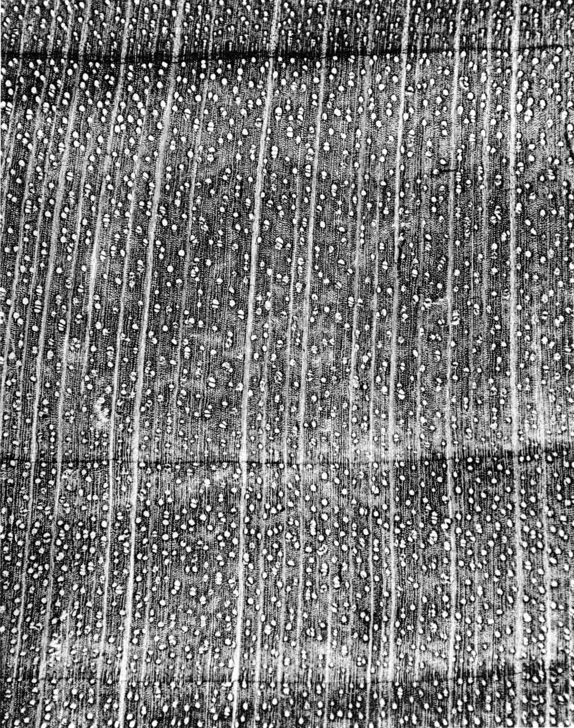
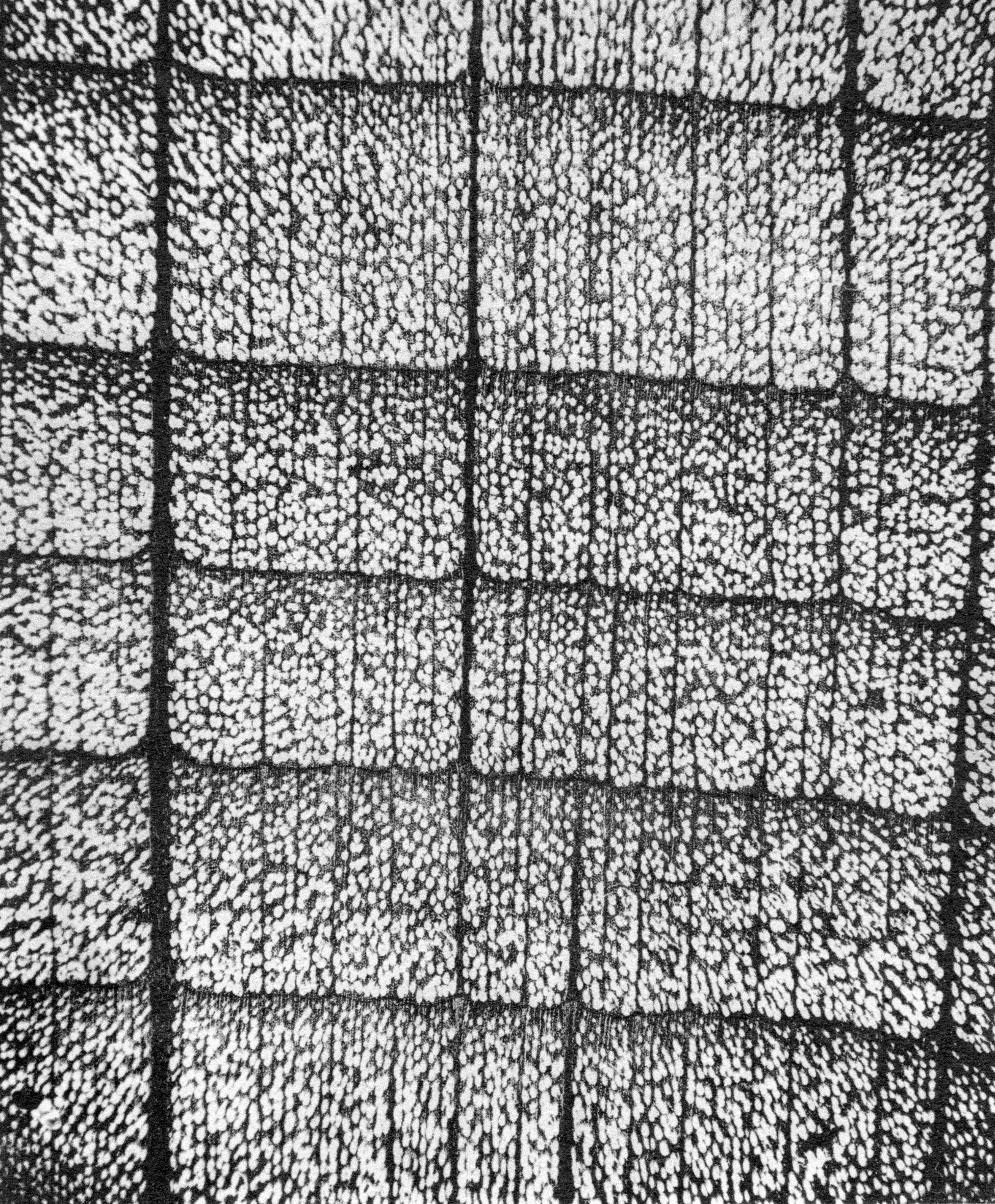
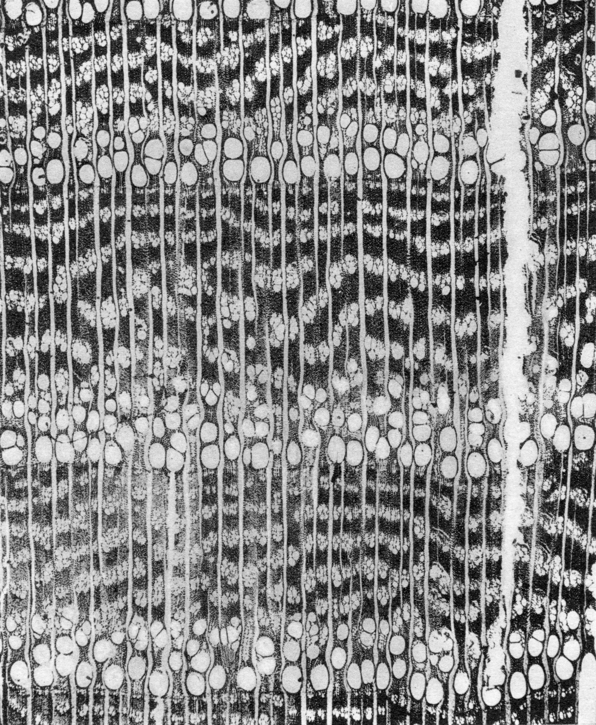
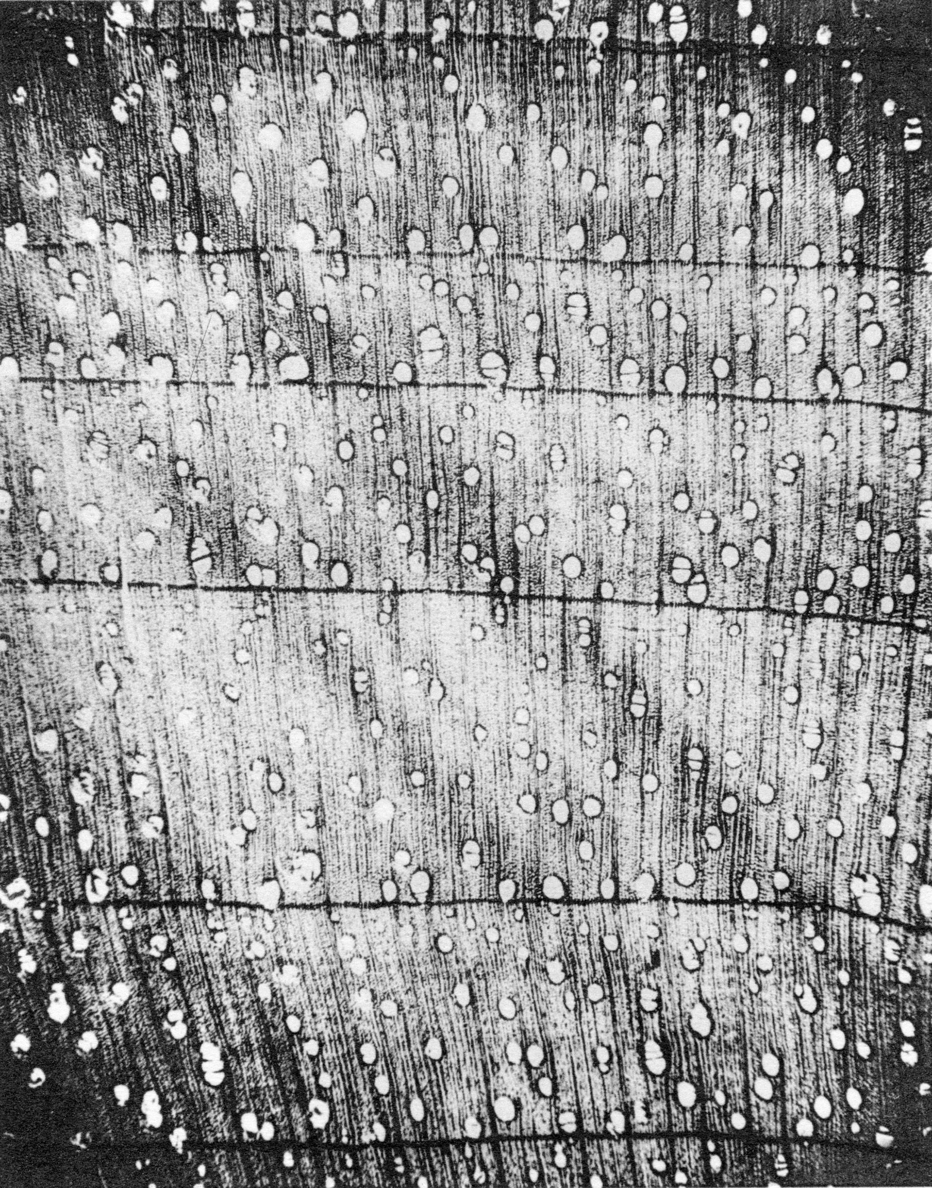
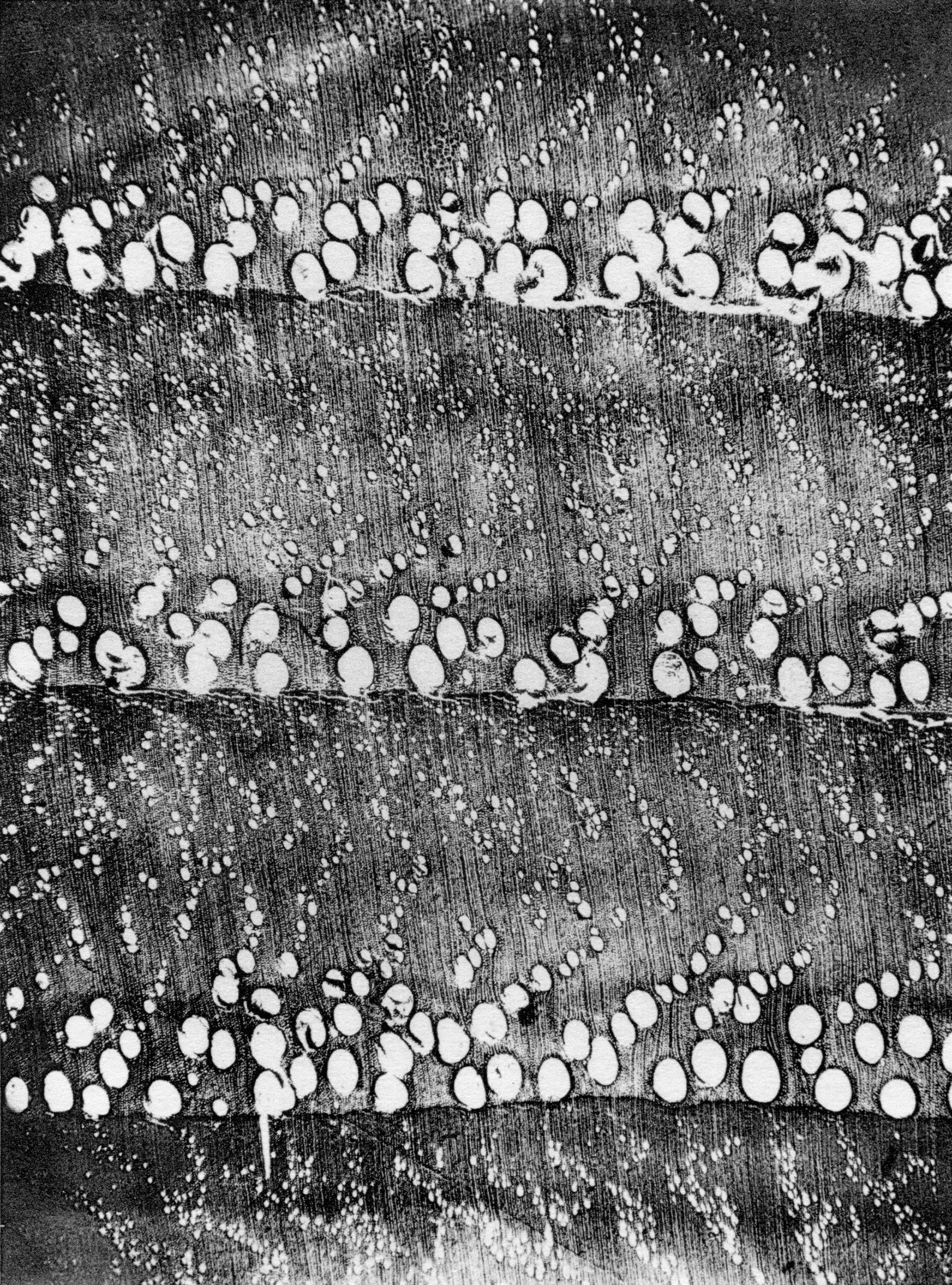
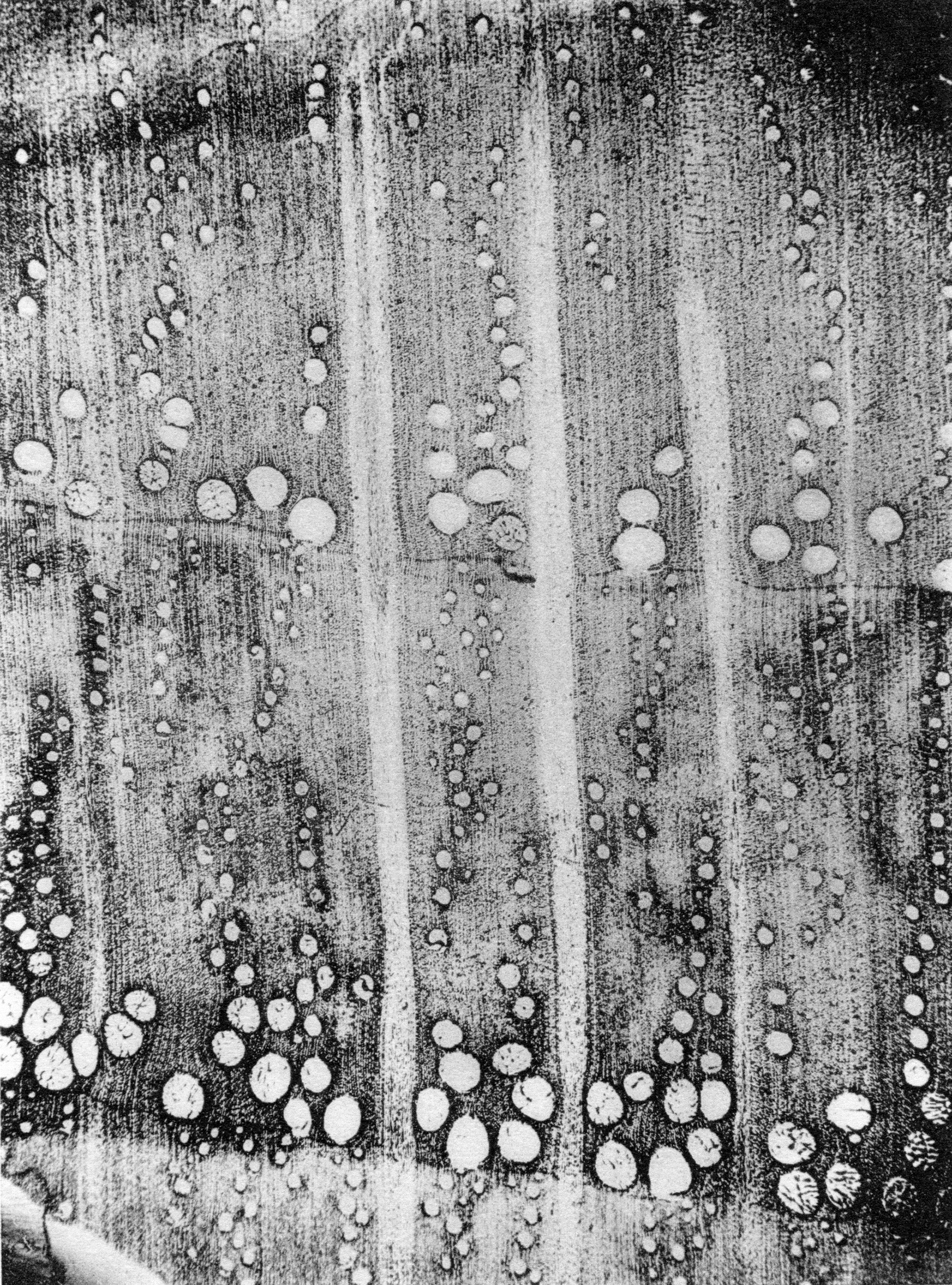
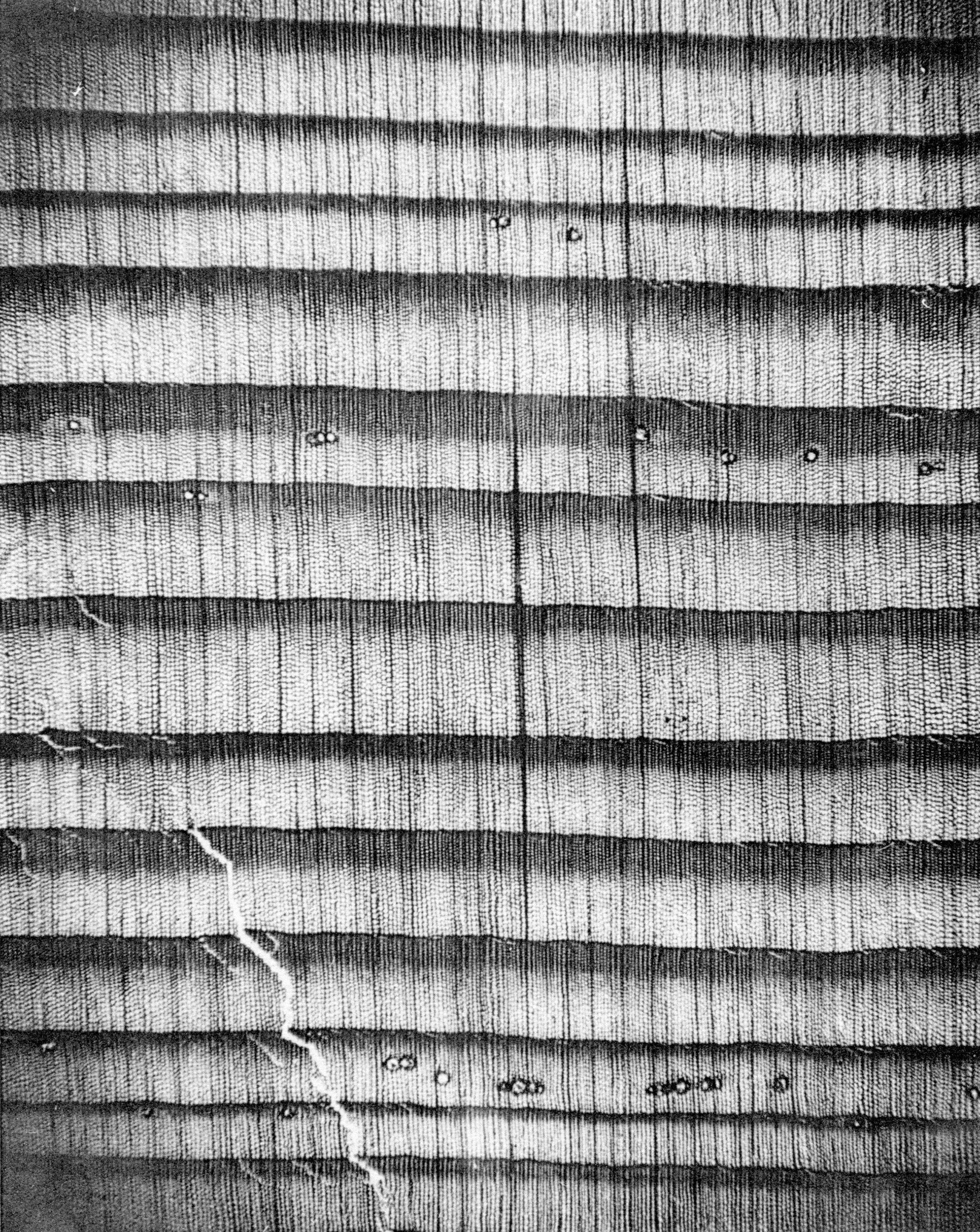
The following microphotographs (Figs 1–15) show the structure of the wood, viewed in cross-section under the microscope. The same characteristics can be observed with a magnifying glass of the same magnification, in reflected light, on smooth-planed crosscut surfaces.
Notes on the identification of different oak and poplar species
The main varieties of European oak can be broken down into two groups of species: deciduous oaks and evergreen oaks. 6Zhang 1997: 63–72. The first group includes sessile, pedunculate, downy, Turkey, and Pyrenean oaks, the first three of which are indistinguishable from one another. Like all deciduous oaks, they form a porous zone at the beginning of each annual growth cycle, comprising pores in the early or spring wood, the diameter of which can reach 500–600 micrometres (µm7µm = 1 thousandth of a millimetre.), and which are hence clearly visible to the naked eye. 8Haneca et al. 2006: 137–44. The second group includes the cork oak (Quercus suber) and holm oak (Quercus ilex). The wood of evergreen oaks lacks this porous zone, and can be readily distinguished from that of deciduous oaks, 9Their very dense and very hard wood has the disadvantage of pronounced shrinkage, which limits its scope for use, although it is very attractive. provided a sufficiently large sample is available, which was not the case with the material we examined. The same applies to poplars: the different species of this tree cannot be distinguished under the microscope. 10The same applies to linden wood and most species.
Figure 2 Willow: hardwood with numerous small and evenly spaced vessels, and very fine rays that do not exhibit figure (10×).
Figure 3 Lime: homogeneous hardwood with fairly numerous, evenly spaced vessels, and relatively thin rays that exhibit fine figure (10×).
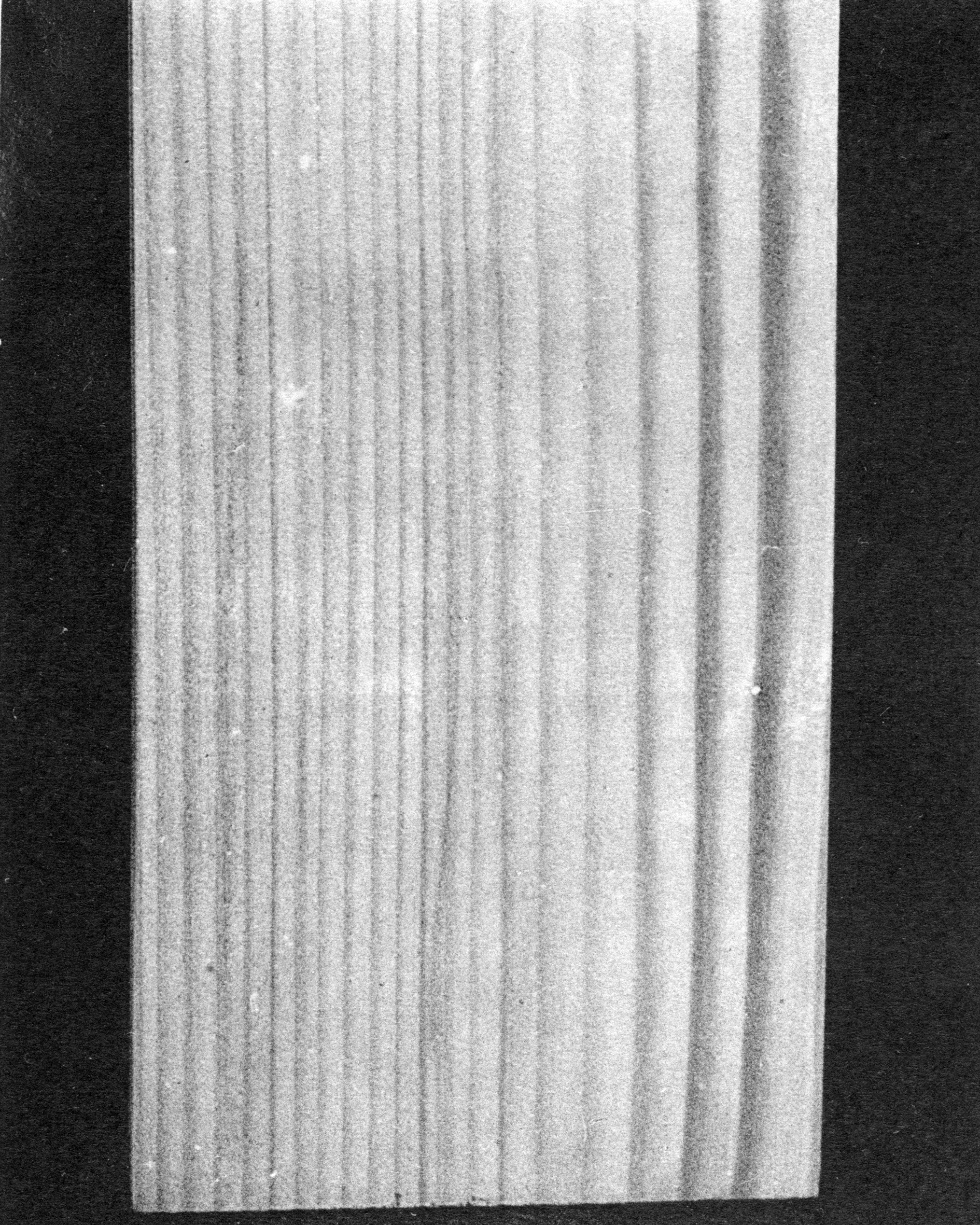
Figure 5 Poplar: hardwood with numerous small and evenly spaced vessels, and very fine, homogeneous rays that do not exhibit figure.
Figure 8 Pear: homogeneous hardwood with numerous vessels and fine rays exhibiting fine figure on a radial cut (50×).
Figure 9 Maple: homogeneous hardwood with fairly numerous, evenly spaced vessels, fairly fine rays, fine figure.
Figure 12 Walnut: homogeneous hardwood with large, scattered vessels, even, fairly narrow rays that exhibit fine figure.
Figure 13 Chestnut: ring-porous hardwood. Vessels of the late wood grouped into radiating flames. Vessels generally elliptical in cross-section. Uniseriate rays, very fine, invisible to the naked eye, hence the absence of figure in radial cut.
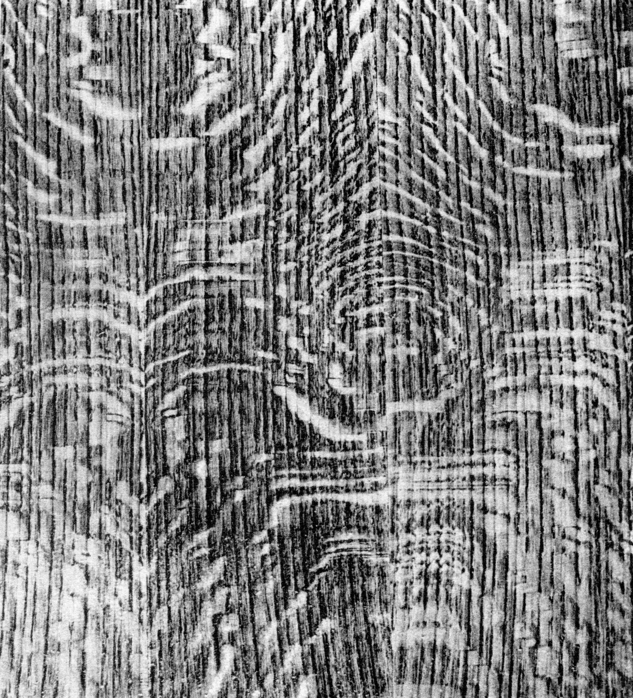
Figure 14 Deciduous oak: ring-porous hardwood, small vessels in flame pattern and some large rays among very fine rays, hence the presence of figure in radial cut.
Fibre and cloth observation techniques
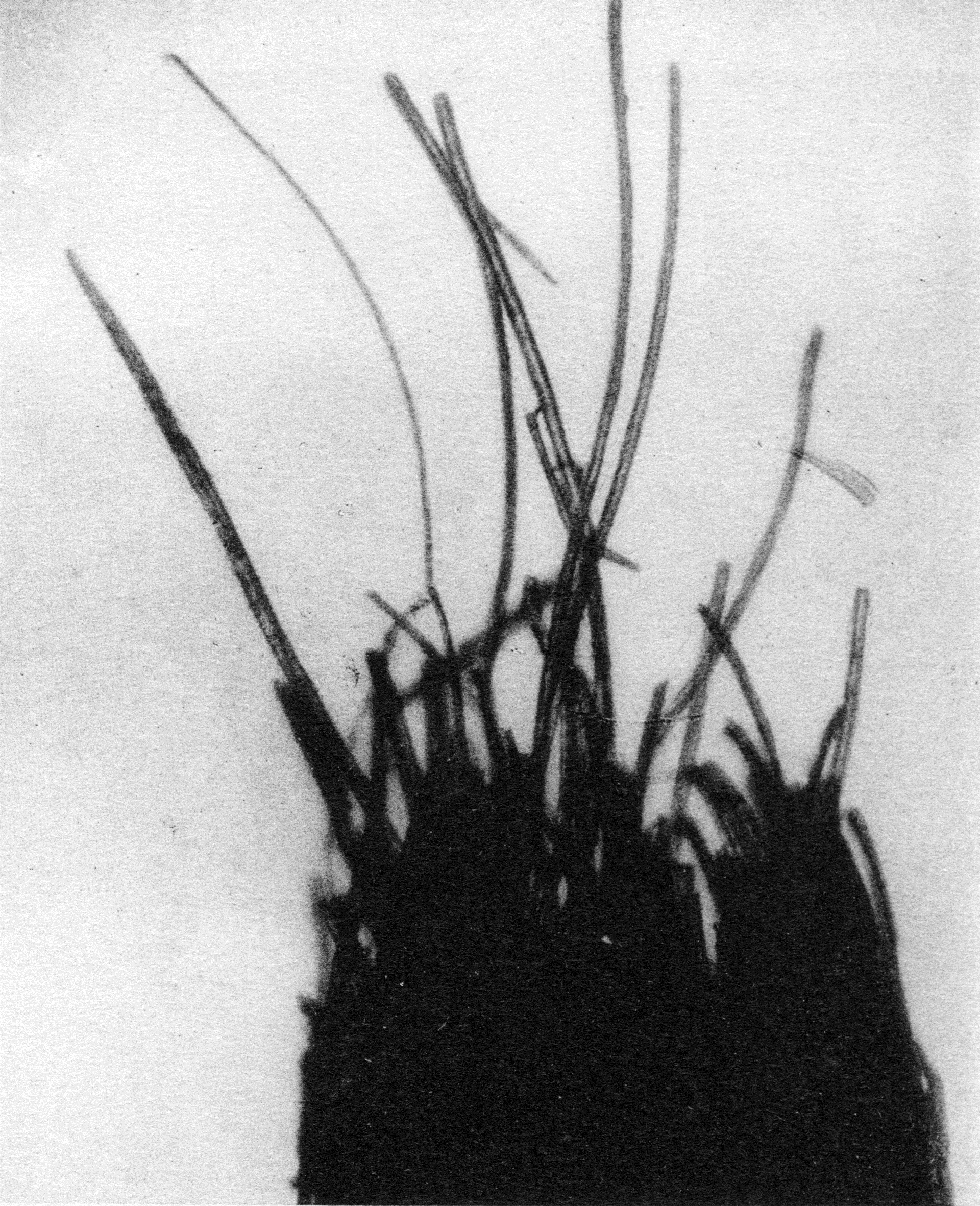
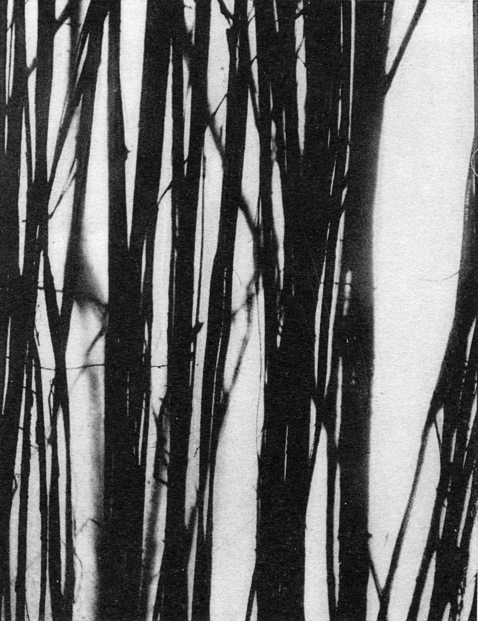
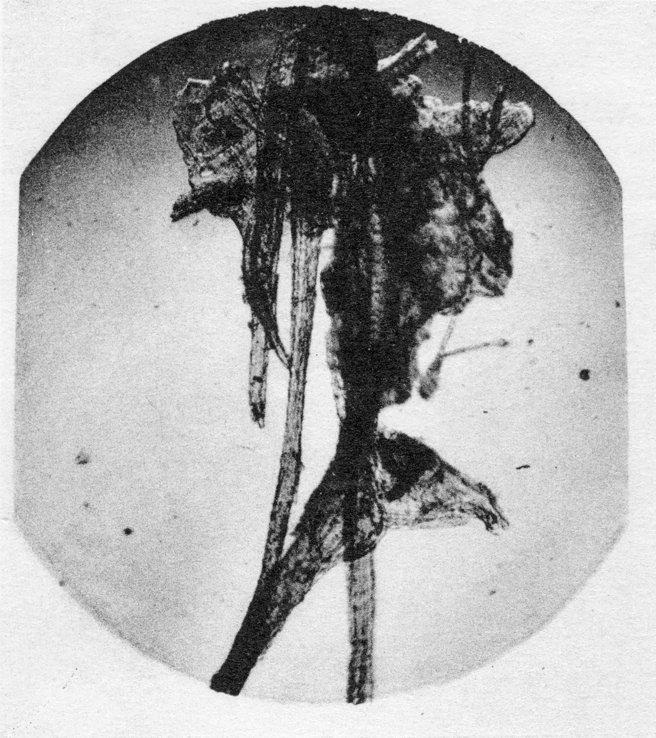
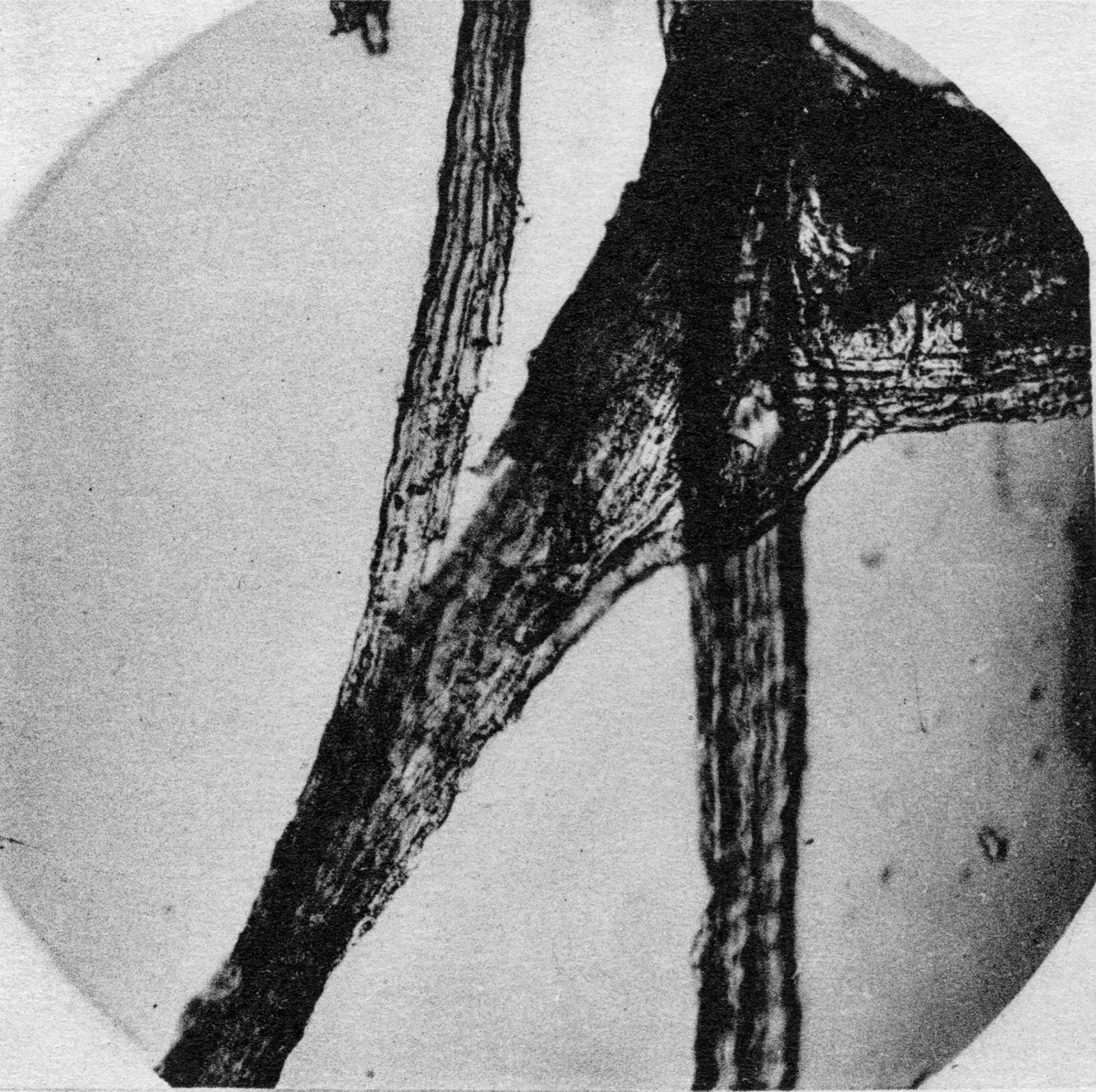
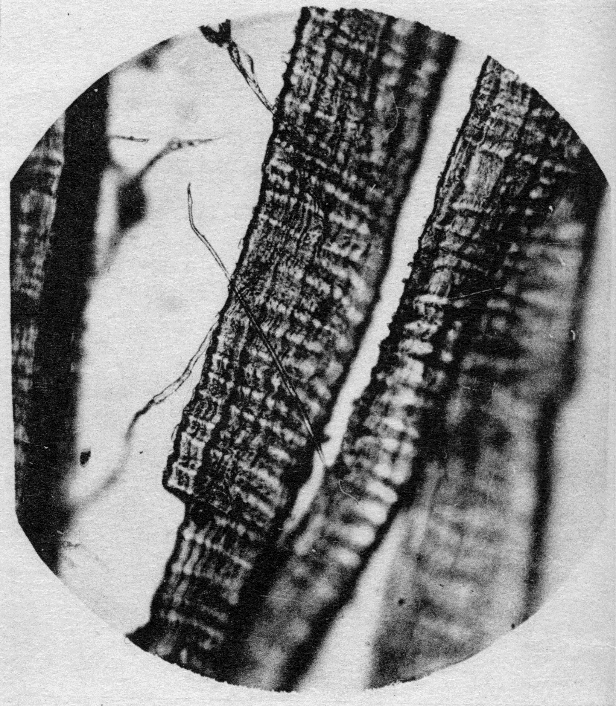
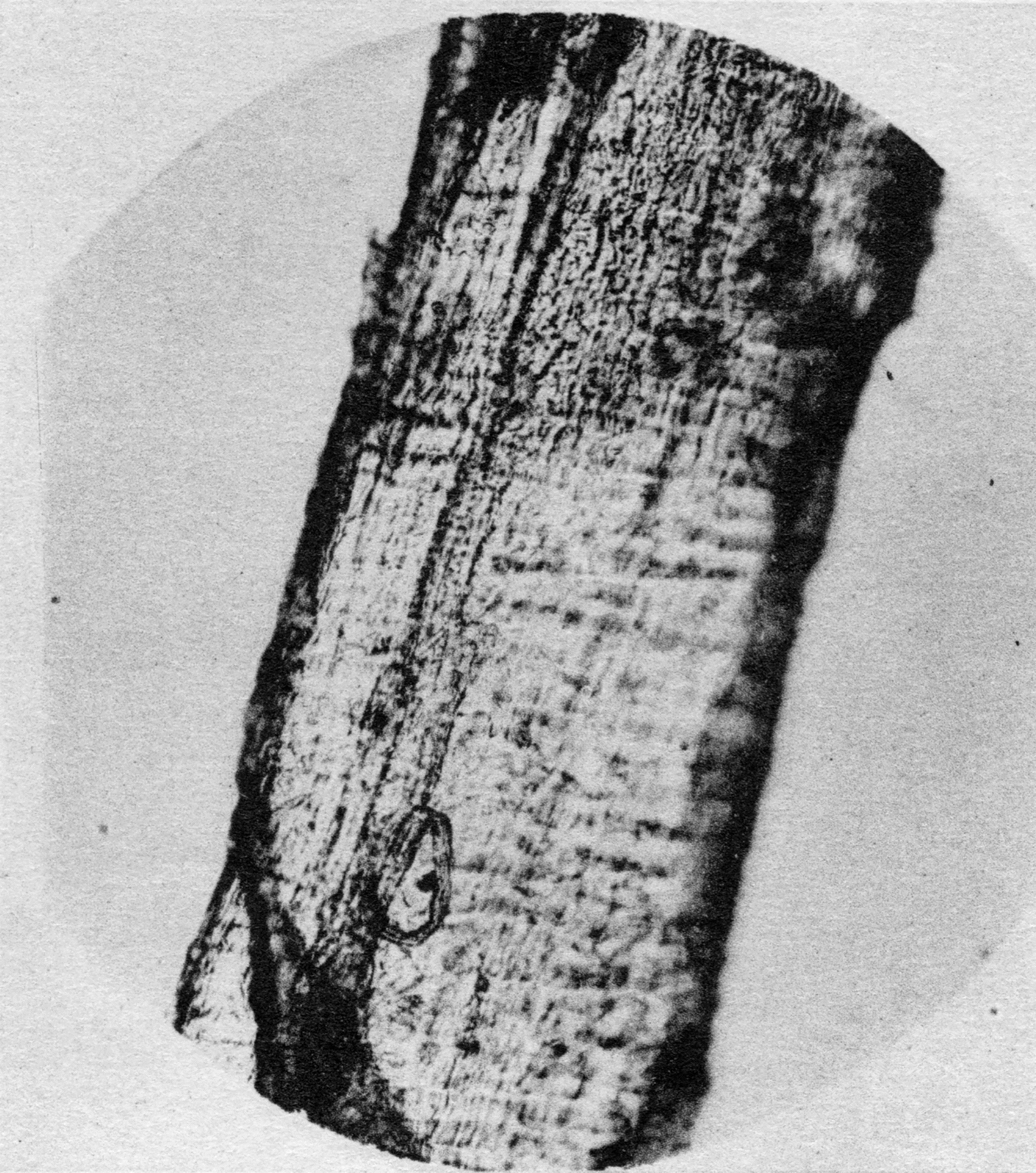
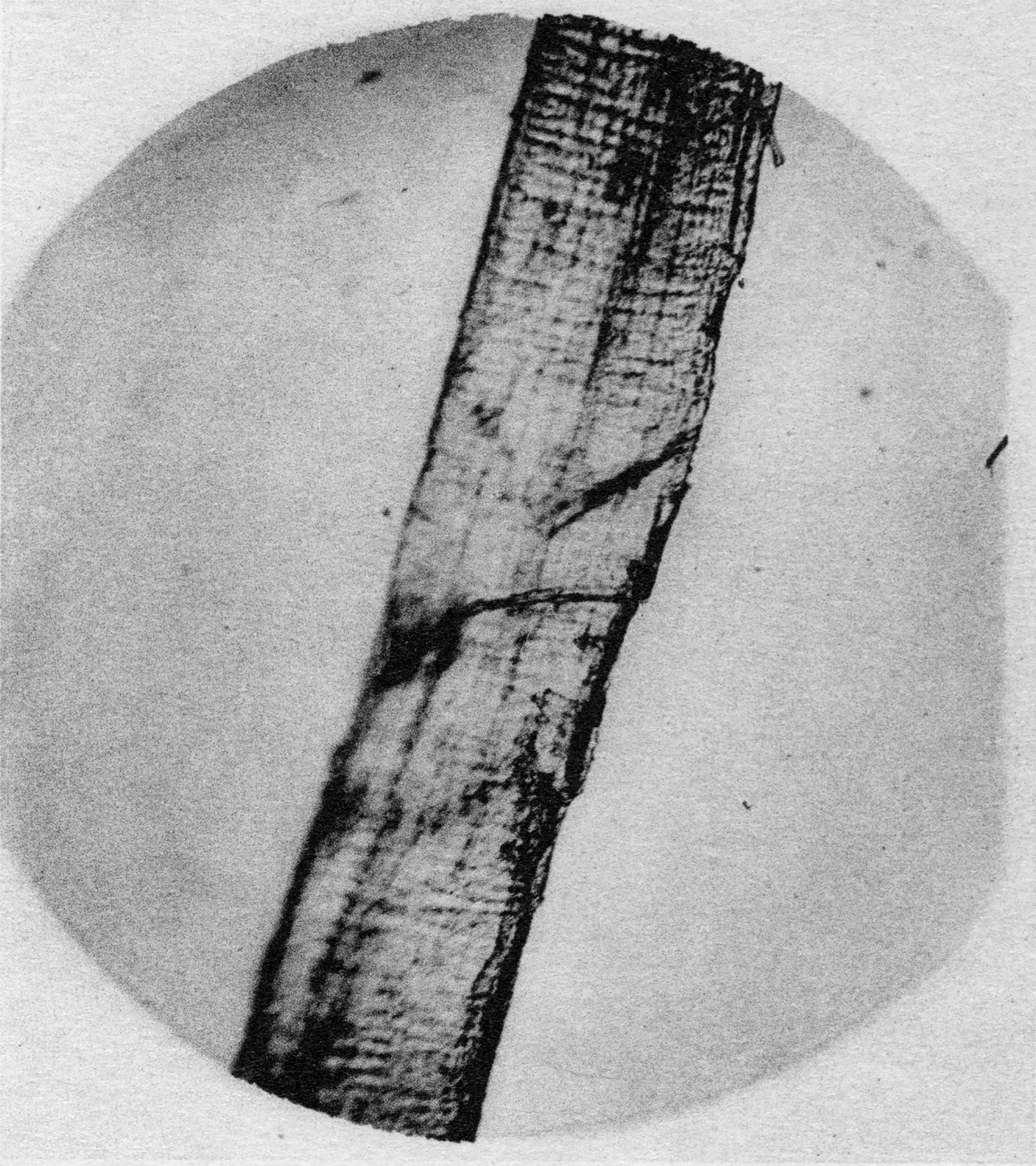
Microscopy, 11Magnification by 50×, 100×, 200×, 400×, corresponding with lenses 5, 10, 20 and 40. which can be used to study the structure of the wood, also enables us to distinguish the nature of cloth and fibre. 12Analysis performed by O. Roherich, Honorary Deputy Director of the Laboratoire de Filature et Tissage at the Conservatoire National des Arts et Métiers. Examination of the fibres revealed the presence of vegetable matter (Figs 16 and 17) and in some cases animal matter (Figs 18–22). The plant matter consisted mostly of hemp fibres and the animal matter of horsehair. 13Skaug 2006: 182–201; Bünsche 2011: 46–60.
X-ray crystallography
X-ray identification
X-ray crystallography 14Analysis performed by Professor A. Guinier of the Sorbonne, at the Laboratoire des Rayons X at the Conservatoire des Arts et Métiers (1961). enables us to identify the crystallized phases present in the preparatory layers, exploiting the propensity of these crystals to diffract X-rays. The graph resulting from this analysis is specific to the type of crystal in question. Examples of the results obtained using this method are presented below (Figs 23–29). The results we have obtained in this way – supplemented in certain cases by chemical analysis – are sufficient to identify the composition of the preparatory layers on the front and back of the panels.
Figure 23 Anhydrite (highly concentrated) + gypsum (concentrated). (Fra Angelico, Angel in Adoration, Paris, Musée du Louvre. Painting index, no. 577.)
Figure 24 Gypsum (highly concentrated) + anhydrite (concentrated, less than in Fig. 23). (Pietro da Rimini, The Descent from the Cross, Paris, Musée du Louvre. Painting index, no. 706.)
Figure 25 Gypsum (highly concentrated) + anhydrite (highly concentrated). (18th-century French School, Barnabas Altarpiece, Paris, Gangnat Collection. Exhib. Paris 1952–53, no. 1; RASAS, Paris 1953, no. 26.)
Figure 26 Anhydrite (highly concentrated) + gypsum (low) + calcium sulphate hemihydrates. (Master of Riofrió, The Assumption, Paris, Musée de Cluny. Painting index, no. 832.)
Figure 27 Calcium carbonate + a little quartz + traces of anhydrite. (Giulio Romano, Portrait of a Man, Paris, Musée du Louvre. Painting index, no. 546.)
Figure 28 Calcium carbonate. (Cimabue, The Virgin and Child with Angels, Paris, Musée du Louvre. Painting index, no. 564.)
Figure 29 Calcium carbonate + traces of quartz. (Dijon workshop, Pietà, Paris, Musée du Louvre. Painting index, no. 175.)
1 Source of the documents on wood anatomy: this chapter and the one that follows were written using information and documents from the Laboratoire d’Anatomie at the Centre Technique du Bois, made available to us by C. Jacquiot, Research Director and Head of the Biology Department. »
2 The science of dendrochronology is part of current methodology in panel painting research. See Klein 1998a; 1998b, 2012; Daly 2007; Fraiture 2007, 2009; Bill et al. 2012. »
4 Klein 2012: 51–65. »
5 More detailed analysis requires use of a microscope. Microscopic examination begins at a magnification of 40, rising as high as 800 for the details. »
6 Zhang 1997: 63–72. »
7 µm = 1 thousandth of a millimetre. »
9 Their very dense and very hard wood has the disadvantage of pronounced shrinkage, which limits its scope for use, although it is very attractive. »
10 The same applies to linden wood and most species. »
11 Magnification by 50×, 100×, 200×, 400×, corresponding with lenses 5, 10, 20 and 40. »
12 Analysis performed by O. Roherich, Honorary Deputy Director of the Laboratoire de Filature et Tissage at the Conservatoire National des Arts et Métiers. »
13 Skaug 2006: 182–201; Bünsche 2011: 46–60. »
14 Analysis performed by Professor A. Guinier of the Sorbonne, at the Laboratoire des Rayons X at the Conservatoire des Arts et Métiers (1961). »
